- Menu Close
- Consumer Goods
- Healthcare
- Performance Materials
-
Technology
- Overview
- Industries
- DKSH Vietnam products
Our products
Search our product database.
-
Services
- Overview
- Sourcing
Sourcing
Accessing a global sourcing network.
- Market insights
Market insights
Generating ideas for growth.
- Marketing and sales
Marketing and sales
Opening up new revenue opportunities.
- Distribution and logistics
Distribution and logistics
Delivering what you need, when you need it, where you need it.
- After-sales services
After-sales services
Servicing throughout the entire lifespan of your product.
- Procurement Transformation Services
Procurement Transformation Services
We support our clients across the entire sourcing lifecycle from sourcing strategy, vendor management, provider selection, supply risk management, implementation, and governance.
- Insights
- Home
- Technology
- DKSH Vietnam products
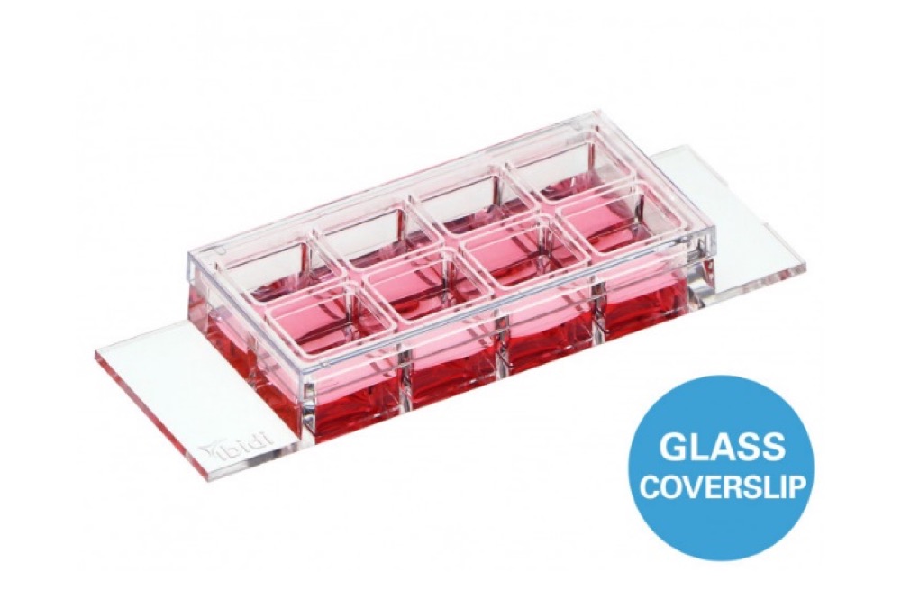
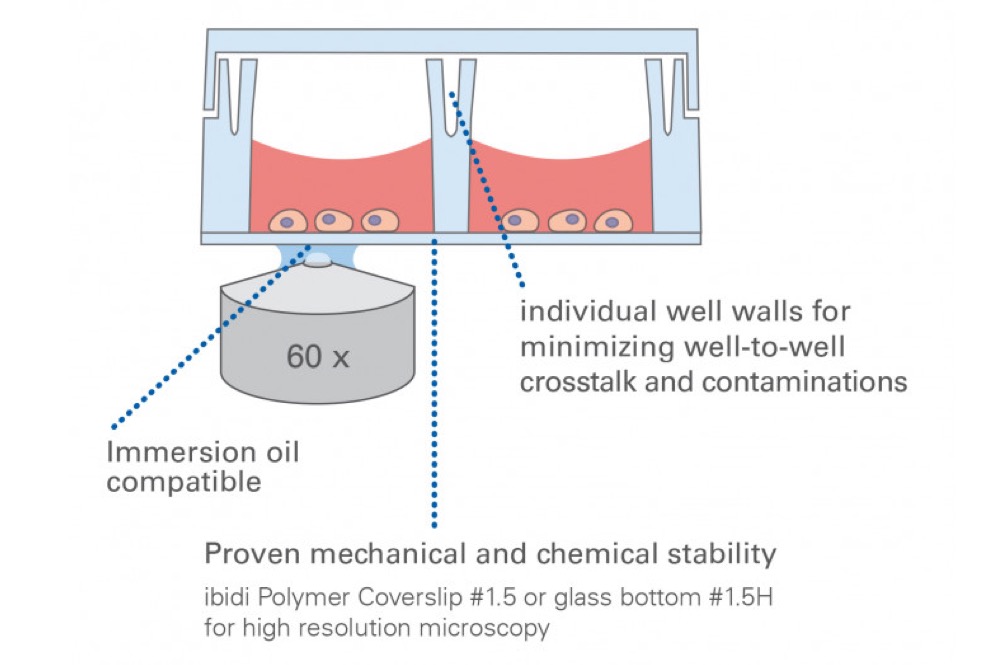
- Ibidi - Labware - µ-Slide 8 Well high
- Home
- Technology
- DKSH Vietnam products
- Ibidi - Labware - µ-Slide 8 Well high