Need support?

Please leave a message

×

Compact digital microscope.

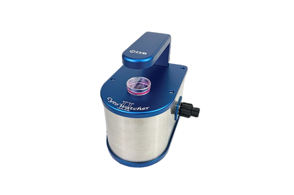
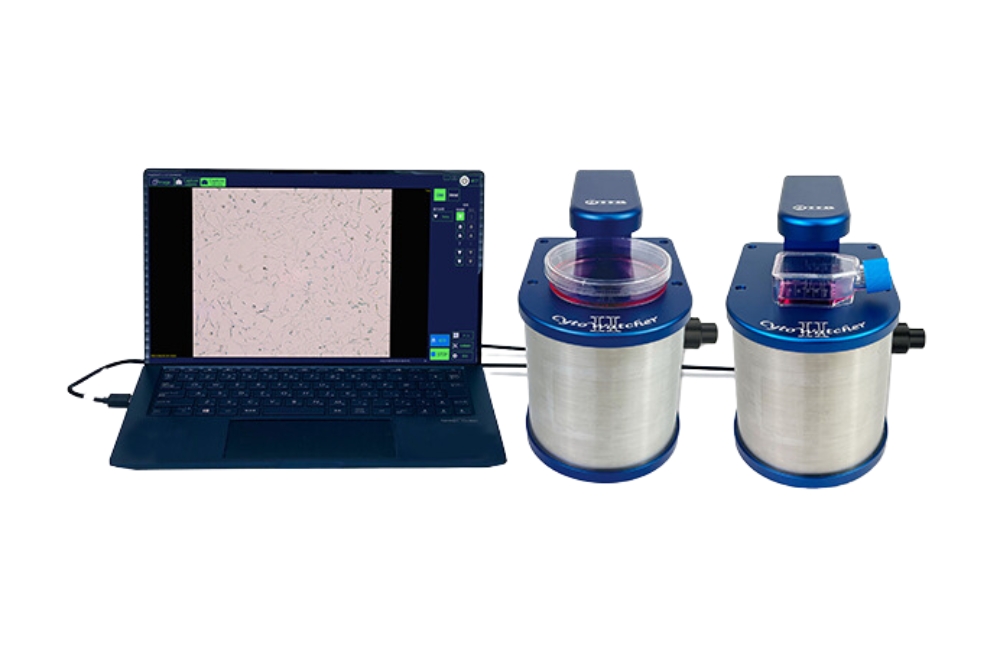
The WSL-1800 CytoWatcher is a digital microscope equipped with a 5-megapixel color CMOS camera. It is designed to facilitate time-lapse live cell imaging with ease. This microscope boasts a space-saving design, making it ideal for laboratories with limited space. Its moisture-resistant construction allows for placement in a CO2 incubator, enabling automated time-lapse imaging while culturing cells. Simply connect the CytoWatcher to a Windows PC using a USB cable to capture and save microscopic images effortlessly.

Application

  • For bright field cell imaging with low magnification.
  • Compact size, no stress to take space.
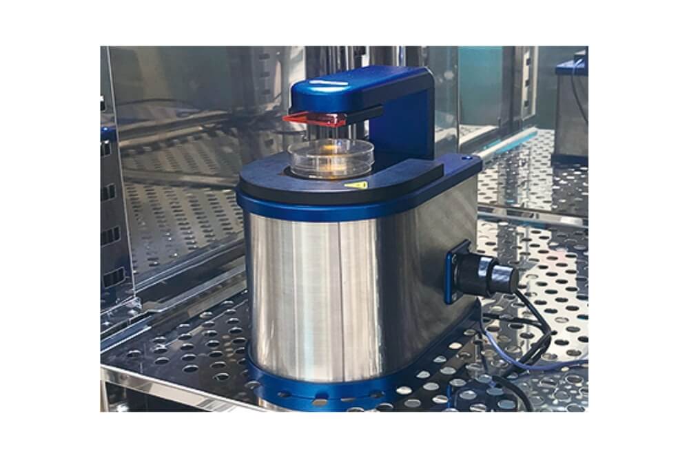
  • Usable in CO2 incubator, for time-lapse imaging in culture condition.
  • Equipped with 5M pixels color CMOS camera.
  • Multiple samples can be taken by connecting multiple units.
  • A model for fluorescent imaging has appeared.

Contact us

Technical Specifications

Model WSL-1850 CytoWatcher I WSL-1850-B CytoWatcher I FL
Camera 5 mega pixels / Color CMOS camera (Global shutter method) 5 mega pixels / Color CMOS camera (Global shutter method)
Resolution 2448 × 2048 pixels 2448 × 2048 pixels
Magnification 4x (Digital zoom : ~ 16x) 4x (Digital zoom : ~ 16x)
Field of view size 1.720 mm x 1.439 mm 1.720 mm x 1.439 mm
Focus Manual (coarse/fine movement) Manual (coarse/fine movement)
Light source White LED (trans-illumination) White LED (trans-illumination)
Side illuminating epi-blue LED (465 nm)
Filter Excitation: 480 nm shortpass filter
Emission: 525 / 45 nm bandpass filter
Moisture resistance Available at 95%H of humidity (Usable in CO2 incubator.) Available at 95%H of humidity (Usable in CO2 incubator.)
Software ImageSaverTmageSaverT for Windows
Capture mode: Live / Time-lapse
Save format: 8bit TIFF / BMP / JPEG, Color / Monochrome
ImageSaverTmageSaverT for Windows
Capture mode: Live / Time-lapse
Save format: 8bit TIFF / BMP / JPEG, Color / Monochrome
Connection to PC USB 3.0 x 1 USB 3.0 Type-A x1 and USB 2.0 (or 3.0) x1
OS Windows 10 / 11 (64/32bit) Windows 10 / 11 (64/32bit)
Language Japanese/English Switchabe with ImageSaverT software Japanese/English Switchabe with ImageSaverT software
Size, Weight 130(W) x 180 (D) x 190(H) mm, 2.5 kg 130(W) x 180(D) x 190(H) mm, 2.9 kg
Power source USB feeding (bus power), DC5V, 7W USB feeding (bus power), DC5V, 7W

Key Features

Since CytoWatcher body is moisture-proof, it can take time-lapsed images of live cells with cultured in a high humidity CO2 incubator. There is no need to use an expensive microscope stage culture system.

  • Compact Design: CytoWatcher is small and lightweight, making it suitable for use in limited spaces such as laboratory benches or safety cabinets.
  • Portability: Easy to carry, allowing you to transport it to your workspace and use it wherever needed.
  • Simple Connection: CytoWatcher connects to a Windows PC using a USB 3.0 cable (optional), with power supplied directly from the PC via USB.
  • Immediate Image Viewing: By launching the control software ImageSaverT (included as standard), users can instantly view live cell images.
  • Easy Image Management: The software allows for easy saving of images and capturing of time-lapse sequences.
  • Multiple Connection Capability: Users can connect multiple CytoWatchers to a single PC, allowing for simultaneous time-lapse imaging of multiple samples in parallel.
  • Up to 2 Units: The system supports connection of up to 2 CytoWatcher units to the PC for increased imaging capacity.
  • Fluorescent Imaging Capability: The fluorescent imaging model features a blue LED light source and excitation/emission filters, supporting green emitted fluorescent imaging such as GFP.
  • Versatile Setup: It can be conveniently set up in a CO2 incubator.
  • Combined Imaging: Capable of combining bright field and fluorescent time-lapse imaging for comprehensive observation.

How to use WSL-1850 CytoWatcher

CytoWatcherⅡ/CytoWatcherⅡ FL is a digital microscope that allows for the long-term imaging of live cells and tissues without causing damage. Designed to be space-efficient and energy-saving, it fits compactly inside a CO2 incubator, minimizing the impact on environmental temperature. It supports not only bright-field imaging but also fluorescence imaging, and its outstanding color reproduction is suitable for observing tissue sections and living tissues.

  • Biopharmaceutical and Biotechnology
  • Education & Academics
  • Medical
  • Pharmaceutical
ATTOATTO Corporation is a scientific instrument developer and manufacturer of protein/nucleic acid research. Long been providing more than 60 years for Polyacrylamide Gel Electrophoresis Devices, PAGEL (precast gel), Western blot solutions, Peristaltic Pumps, Printgraph (High-end Gel Documentation System), Luminograph series (Imaging System for bio/chemiluminescence detection), etc. Their products have been used in many laboratories in Japan and other countries.
[]