- Hàng tiêu dùng
- Chăm sóc sức khỏe
- Hóa chất
-
Ngành Kỹ thuật công nghệ
- Overview
- Ngành hàng
- Sản phẩm của DKSH Việt Nam
Sản phẩm của chúng tôi
Tìm kiếm trong danh mục sản phẩm
-
Dịch vụ
- Overview
- Nguồn cung
Nguồn cung
Tiếp cận mạng lưới nguồn cung toàn cầu
- Thấu hiểu thị trường
Thấu hiểu thị trường
Tạo ý tưởng thúc đẩy tăng trưởng
- Marketing và sales
Marketing và sales
Khám phá nguồn doanh thu mới
- Phân phối và hậu cần
Phân phối và hậu cần
Chuyển giao sản phẩm mọi lúc mọi nơi
- Dịch vụ hậu mãi
Dịch vụ hậu mãi
Hỗ trợ trong suốt vòng đời sản phẩm của bạn
- Insights
-
Vietnam
-
EN | VN
-
Search
- Trang chủ
- Ngành Kỹ thuật công nghệ
- Sản phẩm của DKSH Việt Nam
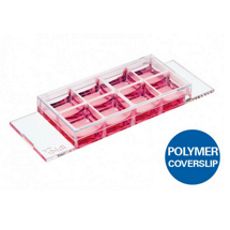
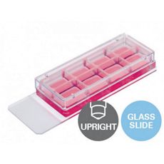
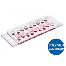
- ibidi – Dụng cụ phòng thí nghiệm – µ-Slide 8 Well High
- Trang chủ
- Ngành Kỹ thuật công nghệ
- Sản phẩm của DKSH Việt Nam
- ibidi – Dụng cụ phòng thí nghiệm – µ-Slide 8 Well High


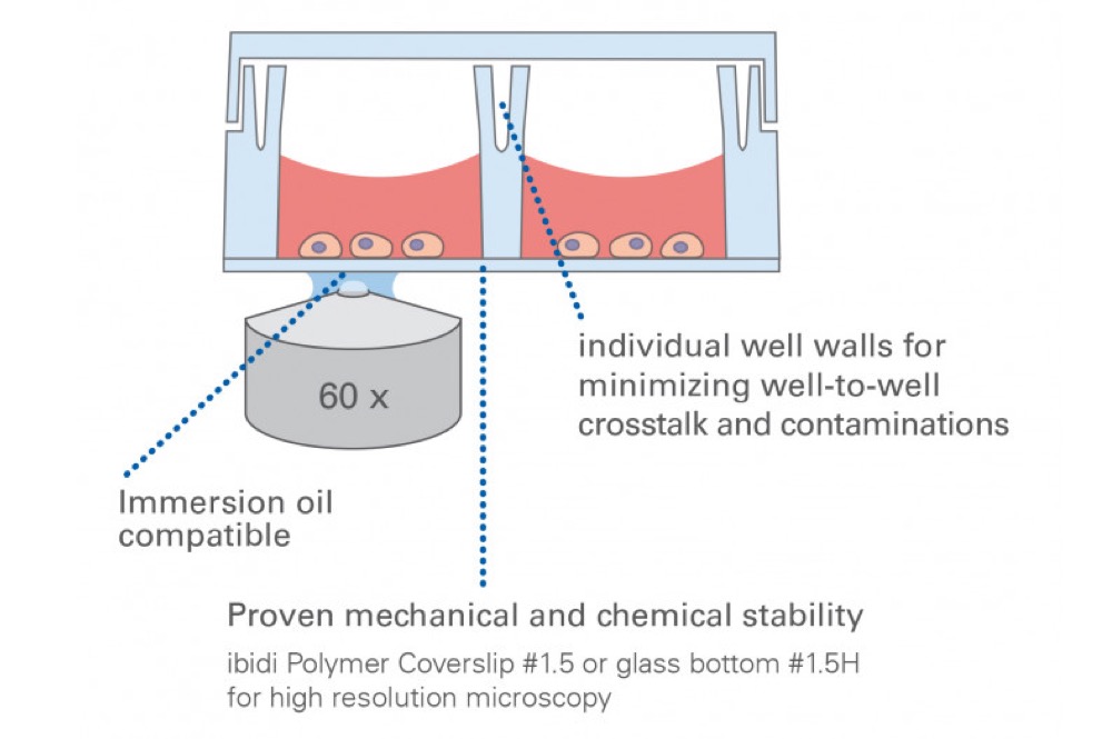


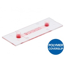
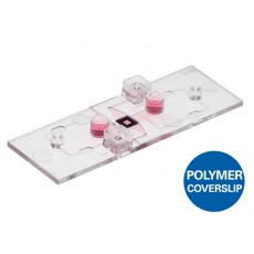
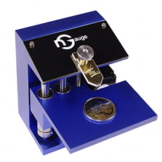
 ibidi - nhà cung cấp dụng cụ xét nghiệm, nuôi cấy tế bào và các sản phẩm tiên tiến cho quá trình sử dụng kính hiển vi tế bào. ibidi với trụ sở chính tại Gräfelfing gần Munich và trụ sở thứ 2 tại Mỹ - Fitchburg, Wisconsin
