- Menu Close
- Hàng tiêu dùng
- Chăm sóc sức khỏe
- Hóa chất
-
Ngành Kỹ thuật công nghệ
- Overview
- Ngành hàng
- Sản phẩm của DKSH Việt Nam
Sản phẩm của chúng tôi
Tìm kiếm trong danh mục sản phẩm
-
Dịch vụ
- Overview
- Nguồn cung
Nguồn cung
Tiếp cận mạng lưới nguồn cung toàn cầu
- Thấu hiểu thị trường
Thấu hiểu thị trường
Tạo ý tưởng thúc đẩy tăng trưởng
- Marketing và sales
Marketing và sales
Khám phá nguồn doanh thu mới
- Phân phối và hậu cần
Phân phối và hậu cần
Chuyển giao sản phẩm mọi lúc mọi nơi
- Dịch vụ hậu mãi
Dịch vụ hậu mãi
Hỗ trợ trong suốt vòng đời sản phẩm của bạn
- Insights
-
Vietnam
-
EN | VN
-
Search
- Trang chủ
- Ngành Kỹ thuật công nghệ
- Sản phẩm của DKSH Việt Nam
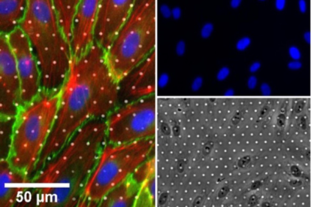
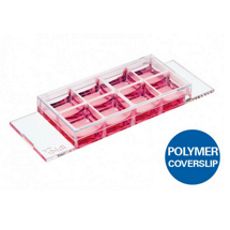
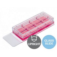
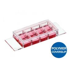
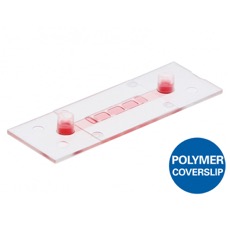
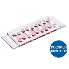
- Ibidi – Dụng cụ phòng thí nghiệm – µ-Slide Membrane ibiPore Flow
- Trang chủ
- Ngành Kỹ thuật công nghệ
- Sản phẩm của DKSH Việt Nam
- Ibidi – Dụng cụ phòng thí nghiệm – µ-Slide Membrane ibiPore Flow