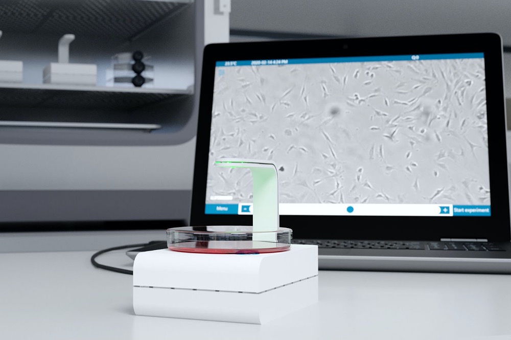
- World’s smallest live-cell imager
Live-cell imaging has become a desired analytical tool in many cell biology laboratories focusing on e.g. pharmacological research, regenerative medicine and tissue engineering. Livecell imaging is generally a difficult task, because it requires large, costly, high-end devices that are difficult to operate. The CytoSMART™ Lux2 is a compact inverted microscope for brightfield live-cell imaging that makes live-cell imaging easy and affordable so it can be used by every biological laboratory. Even in routine cell culture processes.
- No issues with environmental controls
Tight control of the environment (e.g. temperature, CO2) is one of the most critical factors determining the success or failure of a live-cell imaging experiment. When using a conventional microscope equipped with an incubator box, it can be quite a challenge to maintain the cells in a healthy state and functioning normally while being imaged for a longer period of time.
The CytoSMART™ Lux2 operates at lowvoltage and is designed for safe use in a regular CO2-incubator. This enables you to minimize environmental changes, giving you more reliable and repeatable experiments for less work.
- Easy data storage and image analysis
The CytoSMART™ Lux2 can be set to record images at specific intervals (between 5-60 minutes) for minutes, hours and days. In fact, it is one of the few systems that can run for weeks. The recorded images are sent to the CytoSMART™ Cloud where they are analyzed using our custom, cloud-based, image analysis software. You can select the appropriate image analysis algorithm, such as confluence detection, according to the experiment you are performing. The image analysis data is represented in the images as well as graphically in a dashboard.
- Easy access. Anywhere. Anytime.
Thanks to cloud data storage and cloudbased image analysis, you can access your recording and view the cell culture in almost real-time from anywhere on any pc, laptop, tablet or mobile phone with internet access. All the recorded data such as images (.jpg), time-lapse video (.avi) or confluency data (.csv) can be downloaded for further processing. In case you have set a notification, our email alerts will keep you up-to-date on confluence levels or long-term temperature drops.


 CytoSMART is an innovator in kinetic live-cell imaging. Combining compact and fast imaging hardware with powerful image analysis algorithms supported by cloud computing. Automation in time-lapse microscopy and image based cell counting to generate high-quality and robust data.


