- ผลิตภัณฑ์อุปโภคบริโภค
- ผลิตภัณฑ์เพื่อสุขภาพ
- วัตถุดิบอุตสาหกรรม
- เทคโนโลยี
-
บริการของเรา
- ภาพรวม
- การจัดหาแหล่งผลิต
การจัดหาแหล่งผลิต
เพิ่มโอกาสเพื่อเข้าถึงเครือข่ายแหล่งผลิตจากทั่วโลก
- การวิจัยและวิเคราะห์
การวิจัยและวิเคราะห์
เราเพิ่มประสิทธิภาพเพื่อการเติบโตของธุรกิจของคุณ
- การตลาดและการขาย
การตลาดและการขาย
เราเพิ่มโอกาสทางรายได้ให้ธุรกิจของคุณ
- การกระจายสินค้าและโลจิสติกส์
การกระจายสินค้าและโลจิสติกส์
เราจัดส่งสินค้าได้ทุกที่และทุกเวลาที่คุณต้องการ
- การบริการหลังการขาย
การบริการหลังการขาย
เราให้บริการคุณในตลอดช่วงการใช้ผลิตภัณฑ์
- ข้อมูลเชิงลึก
-
Thailand
-
EN | ไทย
-
Search
- กลุ่มดีเคเอสเอช
- ติดต่อ
- ร่วมงานกับเรา
- นักลงทุน
- หน้าแรก
- เทคโนโลยี
- ผลิตภัณฑ์ของเรา
- CytoSMART - Live-Cell Imaging Microscopes - Omni
- หน้าแรก
- เทคโนโลยี
- ผลิตภัณฑ์ของเรา
- CytoSMART - Live-Cell Imaging Microscopes - Omni


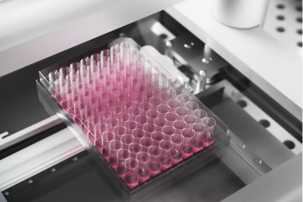
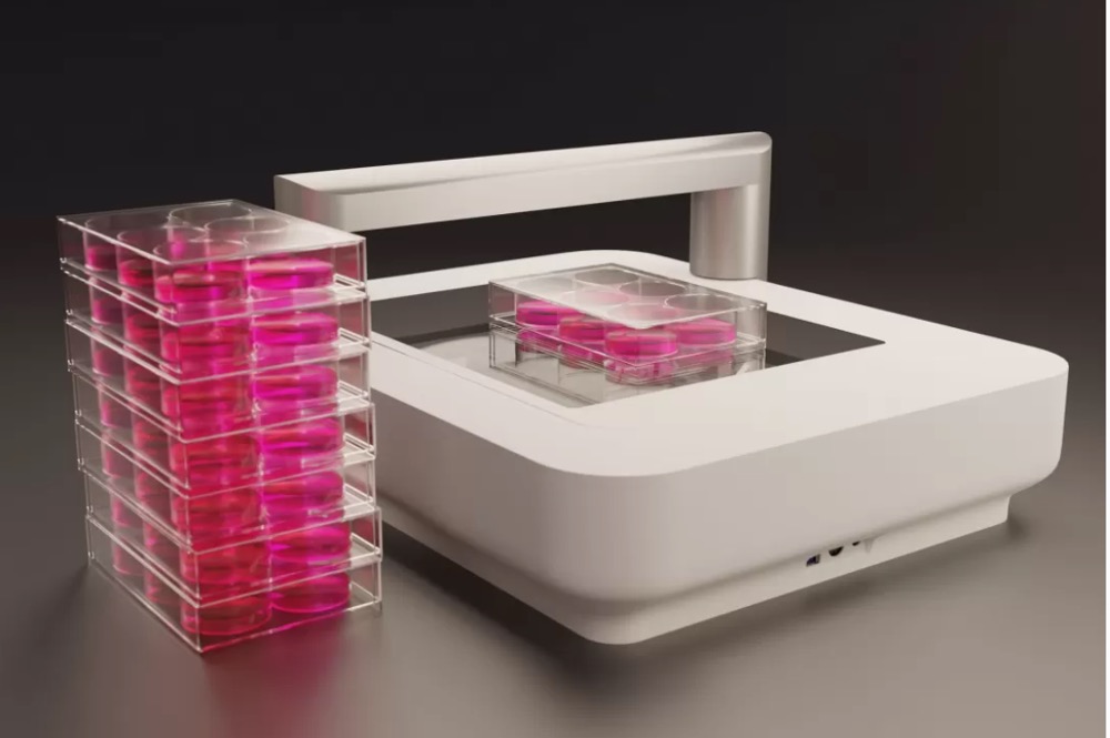
 CytoSMART is an innovator in kinetic live-cell imaging. Combining compact and fast imaging hardware with powerful image analysis algorithms supported by cloud computing. Automation in time-lapse microscopy and image based cell counting to generate high-quality and robust data.


