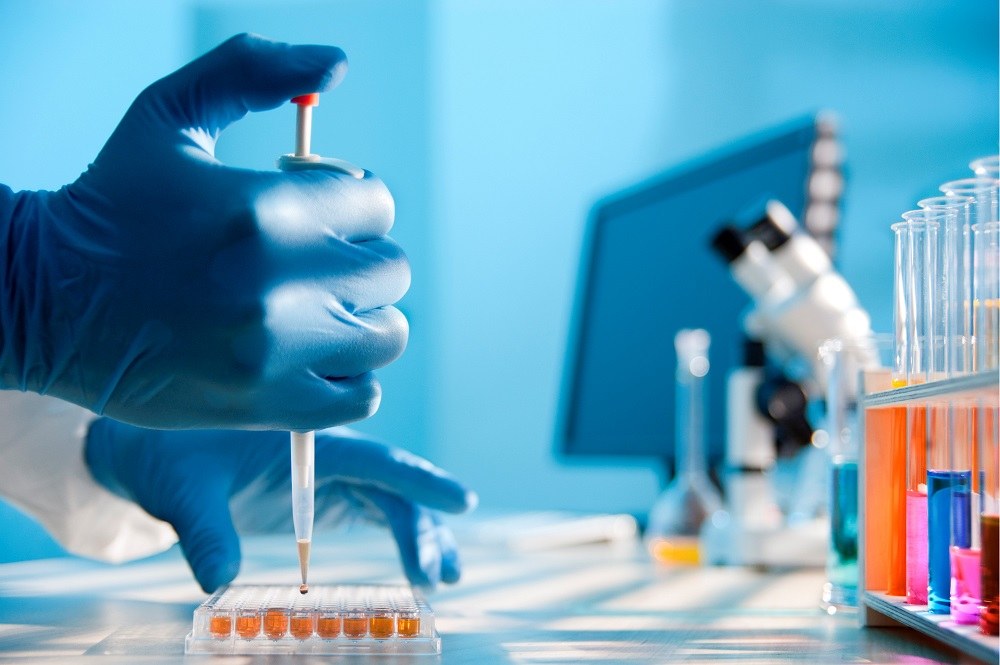
Extracellular vesicles (EV) are nanoparticles found in biological fluids, capable of transporting biological material around the body. They are produced by all cells as part of normal physiological processes and act in cell-to-cell communication.
EVs include exosomes produced and secreted by multivesicular bodies, ectosomes shed from the plasma membrane, and vesicles produced during apoptosis, cell death, and other processes. They carry proteins, nucleic acids, and other molecules from their cell of origin and can deliver this cargo to nearby or distant cells and can be found in various biofluids including plasma, cerebrospinal fluid, urine, and saliva.
There is growing interest in understanding the role of EVs in inter-cellular communication and exploiting them in liquid biopsies for diagnostics purposes, as a therapeutic, and as a vector for drug delivery.
Although the number of research and findings continues to grow, the use of EVs in clinical applications remains limited due to the lack of standardization in EVs isolation and analysis. The two main challenges are heterogeneity and size.
Heterogeneity. EVs can be produced from different places within a cell and are released by many different cell types, EVs found in biofluids are very heterogeneous. As most genomic and proteomic analyses use the bulk analysis method that reports only the population average, the signal from EVs of interest can be lost in the background of irrelevant EVs and particulates of similar sizes.
Small size. Single-particle analysis methods, including flow cytometry, are potentially powerful tools for understanding this heterogeneity, but EVs’ small sizes pose a significant challenge for conventional approaches. Extracellular vesicles range in size from ~50 nm to >1 μm, although the vast majority are between ~70 nm and 200 nm in diameter. As such, they are about 100 times smaller than a typical mammalian cell, with 10,000 times less surface area and 1 million times less volume.
Currently, there is no analytical technique that can detect the entire size range of submicron biological particles with perfect results. Imaging techniques such as electron microscopy offer great resolution, but at very low throughputs. Flow cytometry is uniquely capable of single vesicle analysis of size and heterogeneity, at a relatively high throughput. The principles of flow cytometry are generally applicable to EV analysis as commercial instruments designed to measure small particles become available.
Flow cytometry-based approaches will play an important role in understanding the origins, functions, diagnostic, and therapeutic significance of EVs in health and disease. The key considerations when using fluorescence flow cytometry for EV analysis include the choice of trigger parameters, immunofluorescence, experimental controls, calibration, and standards.
Trigger parameter (light scatter vs fluorescence). In cell analysis, the trigger channel is generally light scatter, usually forward angle light scatters, which allows cells that are much larger than the laser wavelength to be reliably distinguished from debris. However, the vast majority of EVs are smaller than the laser wavelength, which results in much less light scatter, often producing signals smaller than the various sources of background in an instrument.
An alternative approach is the use of fluorescence as a trigger, which can provide improved detection of EVs if the appropriate fluorescence markers are used.
Immunofluorescence. EVs, with their small volume, have very little autofluorescence and sensitivity is limited by various background sources from the sample or instrument. The sample background may result from free dye, which can be decreased by dilution.
Instrument background includes optical background from the scattered laser light and other sources and electronic background from detectors and other components of the data acquisition system. Due to the differences between commercial instruments, and the importance of comparing data between labs and over time, as well as benchmarking new EV analysis systems, appropriate fluorescence calibration is essential.
Experimental controls. EVs used as control experiments are necessary to establish the specificity of single EV detection. Detection thresholds are set near the background levels of the instrument, such that some minimal level of background events will be detected in a buffer-only sample, making this an essential control.
Events being measured are vesicular because samples being analyzed can also contain non-vesicle particulates. A series of two-fold serial dilutions of the stained sample is used for doublet discrimination.
Calibration and standards. Standardization and calibration of EV size estimates, fluorescence measurements of EV cargo, and the confirmation of specificity for measuring single EVs are important and require special attention, especially between instruments.
As the exclusive distributor of extracellular-vesicles compatible flow cytometry solutions like Cytek Biosciences and NanoFCM across various markets in Asia, DKSH is well-positioned to support your business in this increasingly important area of extracellular vesicles research and analysis. Read more at DKSH Lab Solutions.
Sources:
James Hsu joined DKSH in 2019 and is part of the Business Development, Business Unit Technology team in Taiwan. In this role, he is responsible for growing the life sciences and scientific instrumentations business. His previous experience was accumulated in the bustling Asian genomics and proteomics sector, where he worked on bringing a digital PCR startup to market. James graduated from the University of California, San Diego.